FavoriteLoadingAdd to favourites

Microscopes

What are the different types of microscopes?

WHAT WILL YOU LEARN?

  1. What are the different types of Microscopes we can use. 
  2. What type of cells can we see using different types of Microscopes.
  3. When to use different types of Microscopes.
Look at your hands. Check out your skin, your hair and your nails. What if I told you, your hand is made up of millions of tiny structures called cells! In fact, your blood, while liquid, is made up of millions of tiny cells! Don't believe me?

Check this out!

Play Video
Blood is actually made up of many different cells! Red blood cells, white blood cells, platelets and a bunch more! You might be wondering, how can we see all these cells? The answer is Microscopes!

Microscopes make this appear larger. Anything that can only be seen with a microscope is called microscopic. Microscopes allowed scientists to see the building blocks that make up all living things! With microscopes you can see bacteria, cells, viruses and so much more!

Check out the video below 'Our incredible, Microscopic World'.

Scroll over each square below to see under the microscope

Hair

Ant

Strawberry

POllen

In the 1590s Hans and Zaccharias Janssen constructed the first usable microscope.

With modern light microscopes we can magnify an object to 1,500 times it size, but an electron microscope can magnify up to two million times!!! 

  • Scroll over the light microscope to see what an onion looks like under a light microscope. 
  • Scroll over the electron microscope to see inside a cell!

Light Microscope

Electron Microscope

Over time the advancements in microscopy have been tremendous. This has lead to the development of different types of microscope, capable of seeing things we didn’t know existed. There are many different types of microscopes. 

Light microscopes

Most schools have access to light microscopes, so it’s important you know how they work. 

A ‘specimen‘ is what you are looking at, this needs to be very thin to allow light to pass through it. The light travels from the source (usually a lamp) through the specimen and then through a series of lenses. 

This causes the specimen to look much larger than it is! What you see is called a image.

The Stereo microscope, also called a dissecting microscope, has two optical paths at slightly different angles allowing the image to be viewed three-dimensionally under the lenses. Like the monocular microscope, stereo microscopes require a light source, however, unlike a light microscope, the light does not pass through the specimen. This means the specimen doesn’t need to be thinly cut.

The distance between the objective lens and the specimen is much greater than in a monocular microscope, so you can work on your specimen while viewing it. 

The digital microscope, invented in Japan in 1986, uses the power of the computer to view objects not visible to the naked eye.

It connects to a computer monitor via a USB cable, much like connecting a printer or mouse. The computer software allows the monitor to display the magnified specimen. Moving images can be recorded or single images captured in the computer’s memory.

An advantage of digital microscopes is the ability to email images, as well as comfortably watch moving images for long periods.

The Electron Microscope (EM) is a powerful microscope, allowing researchers to view a specimen at nanometer size.

The Transmission Electron Microscope (TEM), the first type of EM, is capable of producing images 1 nanometer in size.

A second type of electron microscope is the Scanning Electron Microscope (SEM) are approximately 10 times less powerful than TEMs, they produce high-resolution, sharp, black and white 3D images.

The TEMs and SEM have practical applications in such fields as biology, chemistry, gemology, metallurgy and industry.

KEY POINTS

  • Monocular microscope require a thin specimen, a light source and produce a 2D image. 

  • Stereo microscopes can produce a 3D image and allow you to work on a live specimen. 

  • Electron microscopes use electrons, not of light. They can magnify specimens to up-to a million times. 

Test your knowledge

Click the ‘question mark’ icon to test your knowledge with some questions.

WHERE NEXT?

Discover more about this topic or go back to the year 7&8 page.

Cell Division

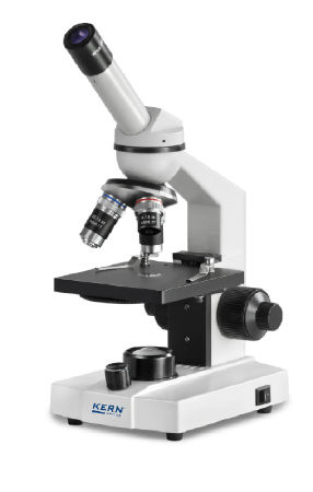
Add to favouritesYear 7 & 8 TOC Specialised cells Cell Division Recognising that cells reproduce via cell division & describing mitosis as cell division for

Read More »

The black plague

Add to favouritesWOW SCIENCE The black plague The black plague, also referred to as the black death, ravaged Europe between 1347 and 1351. During that

Read More »

What are cells?

Add to favouritesYear 7 & 8 TOC Building Blocks Cells Distinguishing plant cells from animal and fungal cells Scientists have identified cells as the basic

Read More »

Wow: Tardigrades

Add to favourites Year 7 & 8 Topics Tardigrade wow science Tardigrades are some of the most toughest organisms on earth! If you think bears,

Read More »

Enjoying our resources?

Everything we provide is free. Help us keep this resource free by donating and helping us cover our running costs. Every little bit helps!